 Espiral Atlas
Lateral malleolus
malleolus lateralis
Study entry
Description
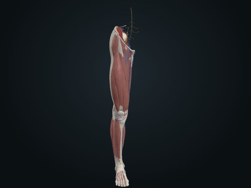
Enlarged distal end, which extends to the side and downward; more prominent and more posterior than the medial malleolus and about 1 cm lower.
Relations
It forms the lateral wall of the ankle mortise; attachment of ligaments of the talocrural joint.
Palpation
Under the skin, with a sharp tip.
Source: cap. 7, «Huesos del miembro inferior: fíbula» y «Anatomía de superficie»
Clinical correlation
Fractures of the fibula
The fibula usually breaks 2 to 6 cm above the tip of the lateral malleolus, often together with a fracture-dislocation of the ankle and a fracture of the tibia. When a person slips and the foot turns in exaggerated inversion, the ligaments of the ankle tear and the talus strikes the lateral malleolus and cuts it off. Fractures of both malleoli are common in soccer and basketball. It hurts because muscle attachments are torn, and walking gets worse because the fibula stabilizes the ankle.
Ankle sprain and fractures of the lateral malleolus
The ankle is the large joint most injured in the body, and the sprain (tearing of ligament fibers) is very common. It is almost always by inversion: the foot, in plantar flexion and bearing load, steps on uneven ground and turns inward. Sprain of the lateral collateral ligament is typical of sports with running and jumping; 70% to 80% of basketball players have had one. The lateral collateral suffers because it is much weaker than the medial and is the one that resists inversion. Its anterior talofibular part is the most vulnerable and the one that tears most, leaving the ankle unstable; the calcaneofibular may also tear.
Related clinical cases
- Fractures of the fibula
- Ankle sprain and fractures of the lateral malleolus