 Espiral Atlas
Teeth
dentes
Study entry
Location
In the dental sockets of the alveolar processes of the maxilla and of the alveolar part of the mandible.
Structure
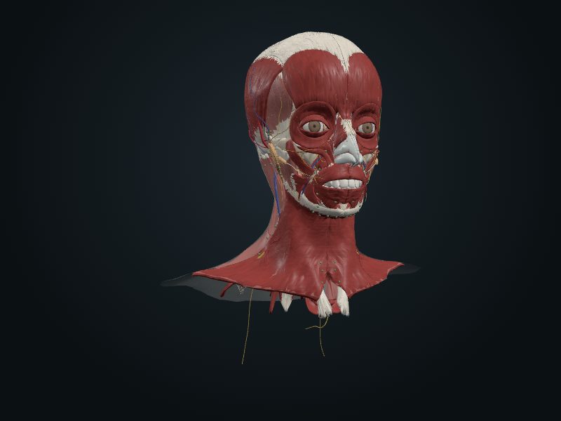
They serve chewing and the articulation of speech. A child has 20 deciduous teeth; an adult, normally 32 permanent ones. Types: incisors (thin, cutting edges), canines (a prominent cone), premolars (two cusps) and molars (three or more). The vestibular surface (labial or buccal) faces outward and the lingual, inward; the mesial surface faces the median plane of the facial part of the skull and the distal moves away from it; both are contact surfaces. Each tooth has a crown (above the gum), neck and root, attached to the socket by the periodontium; the number of roots varies. Most of the tooth is dentin, covered by enamel on the crown and by cementum on the root. The pulp cavity contains connective tissue, vessels and nerves, which enter and leave through the root canal. The sockets are the skeletal structures that change most over life; the interalveolar septa separate the sockets and the interradicular ones, the roots of the same tooth. The labial wall of the socket is very thin in the incisors and the lingual one in the molars; for this reason the labial surface usually breaks to extract the incisors and the lingual one, the molars.
Blood supply
Superior and inferior alveolar arteries, branches of the maxillary; the veins accompany the arteries.
Innervation
Dental plexuses formed by branches of the superior alveolar (CN V2) and inferior alveolar (CN V3) nerves.
Lymphatics
The lymphatic vessels of the teeth and gums drain mainly into the submandibular lymph nodes.
Clinical
Caries (cavities) develop when acids and enzymes of the bacteria of the mouth break down the hard tissues; untreated ones invade the pulp cavity and cause pulpitis.
Source: cap. 8, «Dientes»; tablas 8-13 A e B
Clinical correlation
Gingivitis and periodontitis
Poor oral hygiene leaves food remains and bacteria on the teeth and in the gingival crevices. This inflames the gums (gingivitis), which become swollen and red. Without treatment, the inflammation reaches the other supporting structures, including the alveolar bone, and becomes periodontitis, with destruction of bone and periodontium. Dentoalveolar abscesses, with pus from necrosis of the inflamed tissues, can drain into the oral cavity and the lips.
Caries, pulpitis and dental abscess
Acids and enzymes from bacteria of the mouth destroy the hard tissues of the tooth and form caries. Without treatment, it invades the pulp cavity and inflames the pulp (pulpitis). The pulp cavity is enclosed in a rigid structure, so the swelling hurts a lot. The pressure can destroy the small vessels of the root canal, and the infected material leaves through the apical foramen and forms an abscess in the alveolar bone. The pus from an upper molar can go to the nasal cavity or the maxillary sinus, and the roots of the lower molars lie near the mandibular canal, where the abscess can compress the nerve.
Upper molars and the maxillary sinus
The three upper molars lie very close to the floor of the maxillary sinus. In extraction, a root can break. If it is pushed upward wrongly, it enters the sinus and creates a communication between it and the mouth, with risk of infection. The superior alveolar nerves supply the upper teeth and the mucosa of the sinus. For this reason inflammation of the sinus often gives pain in the molar.
Supernumerary teeth
Supernumerary teeth are those beyond the normal number. They can be single or multiple, unilateral or bilateral, erupted or not, in one or both arches. They appear in both dentitions, more in the permanent. The most common is the mesiodens, peg-shaped, between the upper central incisors. Rarely are there many without another associated condition, such as cleft lip or cleft palate. They can interfere with the eruption and alignment of the normal teeth and are usually removed.
Related clinical cases
- Gingivitis and periodontitis
- Caries, pulpitis and dental abscess
- Upper molars and the maxillary sinus
- Supernumerary teeth