 Espiral Atlas
Tibial tuberosity
tuberositas tibiae
Study entry
Description
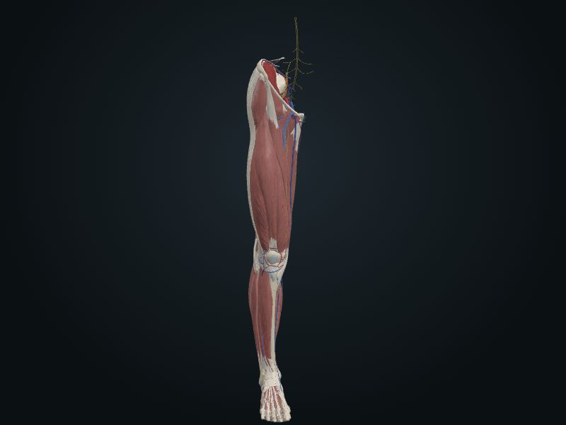
A broad, oval elevation at the top of the anterior border.
Relations
Insertion of the patellar ligament.
Palpation
Easy, about 5 cm below the apex of the patella.
Source: cap. 7, «Huesos del miembro inferior: tibia» y «Anatomía de superficie»
Clinical correlation
Epiphysial plates of the tibia and Osgood-Schlatter disease
The center of ossification of the upper end of the tibia appears soon after birth and fuses with the shaft in adolescence, usually between 16 and 18 years. In a child, a fracture that crosses the epiphysial plate is more serious because it can interfere with growth. The tibial tuberosity usually grows downward from the upper center at about 10 years, but sometimes has a center of its own, near 12. If the plate is torn at the tuberosity, it becomes inflamed and hurts chronically and recurrently in adolescence: this is Osgood-Schlatter disease, typical of the young athlete.
Intraosseous infusion in the tibia
Intraosseous infusion delivers fluids, blood and drugs straight to the medullary cavity when a peripheral vein cannot be obtained, especially in traumatic shock and in a child in circulatory collapse. The most used site is the proximal tibia: the skin is thin and there are landmarks that help place the needle in the marrow without reaching the growth plate. The needle enters the flat area of the bone, about 2 cm below and slightly medial to the tibial tuberosity. Other sites: distal femur, distal tibia or fibula, proximal humerus and manubrium. Because of the risk of osteomyelitis, it is replaced by venous access within 24 hours.
Related clinical cases
- Epiphysial plates of the tibia and Osgood-Schlatter disease
- Intraosseous infusion in the tibia