 Espiral Atlas
Greater trochanter
trochanter major
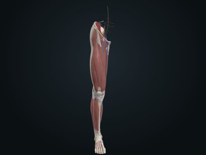
Study entry
Description
A large, lateral bony mass, which rises and goes backward where the neck meets the shaft.
Relations
Insertion and lever of the abductors and rotators of the thigh.
Palpation
On the lateral surface of the thigh, about 10 cm below the iliac crest; the posterior margin is the easiest, with the limb unloaded.
Source: cap. 7, «Huesos del miembro inferior: fémur» y «Anatomía de superficie»
Clinical correlation
Fractures of the femur
Although large and strong, the femur breaks often, and the type of fracture follows age and sex. That of the neck is the most common: it is the narrowest and weakest part and forms an angle with the line of load; it becomes more fragile with age, especially in women with osteoporosis. Proximal fractures (transcervical, intertrochanteric) come from indirect trauma, such as tripping or stepping down hard from a step. They are unstable: the fragments overlap and the limb shortens, helped by muscle spasm. Intracapsular ones also put the femoral head at risk, since it loses its blood supply.
Trochanteric bursitis
The trochanteric bursa becomes inflamed with repeated movements that use the gluteus maximus, such as climbing stairs carrying a load or running on a steeply inclined treadmill: the tendon fibers move back and forth over the bursa, on top of the greater trochanter. It gives deep, diffuse pain on the lateral surface of the thigh, with a tender point over the greater trochanter, and radiates along the iliotibial tract (from the iliac tubercle to the tibia), a thickening of the fascia lata reinforced by the tensor fasciae latae and the gluteus maximus.
Related clinical cases
- Fractures of the femur
- Trochanteric bursitis