 Espiral Atlas
Mandibular central incisor
dens incisivus medialis mandibularis
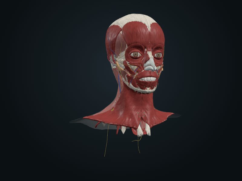
Study entry
Location
Socket of the alveolar part of the mandible, on each side of the mandibular symphysis.
Structure
It is the smallest of the teeth. The crown is narrow and symmetrical, with a straight incisal edge; the root is single and flattened mesiodistally, and can have one or two canals. It erupts at about 6 to 8 years, a little before the upper incisor.
Neighborhood
The two central incisors lie side by side, in the midline, behind the lower lip and in front of the region of the genioglossus muscles.
Blood supply
Inferior alveolar artery, branch of the maxillary, which runs in the mandibular canal and gives a branch to each root; the veins accompany the artery.
Innervation
Inferior dental plexus, formed by the inferior alveolar nerve (CN V3), which runs in the mandibular canal.
Lymphatics
They drain into the submental and submandibular lymph nodes.
Clinical
Dental calculus (tartar) forms easily on the lingual surface of the lower incisors, near the ducts of the submandibular and sublingual glands. They are usually the first permanent teeth to appear, together with the first molar.
Source: cap. 8, «Dientes»; tabla 8-13 B (erupción de los dientes permanentes)