 Espiral Atlas
Maxillary central incisor
dens incisivus medialis maxillaris
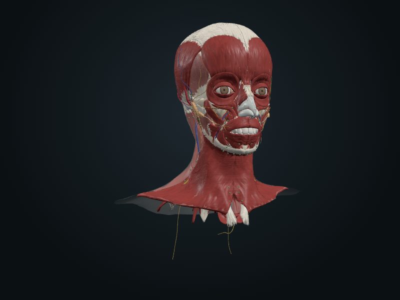
Study entry
Location
Socket of the alveolar process of the maxilla, on each side of the midline, just below the nose.
Structure
It is the largest of the incisors and the most visible when smiling. The crown is wide, shovel-shaped, with a straight incisal edge, smooth vestibular surface and concave palatal surface, with a cingulum (prominence next to the gum) and two marginal ridges. The root is single, conical and straight, with one canal. It erupts at about 7 to 8 years.
Neighborhood
The two central incisors touch by their mesial surfaces, in the midline. The frenulum of the upper lip descends between them to the incisive papilla of the palate.
Blood supply
Anterior superior alveolar artery (branch of the infra-orbital, which comes from the maxillary); the veins accompany the artery.
Innervation
Superior dental plexus, formed by the anterior superior alveolar branch of the infra-orbital nerve (CN V2).
Lymphatics
The lymphatic vessels of the tooth and gum drain mainly into the submandibular lymph nodes.
Clinical
It is the tooth most affected by dental trauma (fall, sport, accident), mainly in children. A thick labial frenulum or an extra tooth (mesiodens) between the central incisors causes a diastema.
Source: cap. 8, «Dientes»; tabla 8-13 B (erupción de los dientes permanentes)