 Espiral Atlas
Mandibular third molar
dens serotinus mandibularis
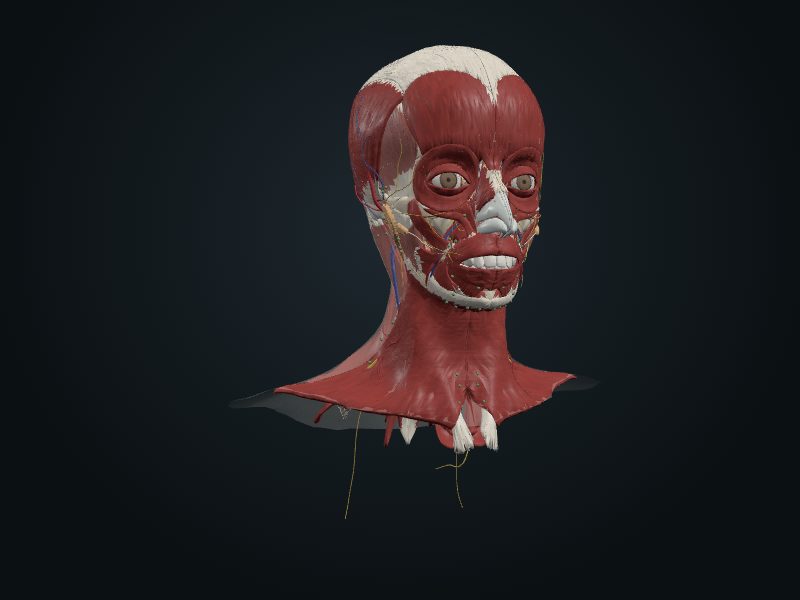
Study entry
Location
Socket of the alveolar part of the mandible, at the end of the arch, next to the ramus of the mandible.
Structure
The last tooth to erupt and the most variable: the crown has three to five cusps and the roots are usually short, curved and fused. It may be absent. The 3D model does not include this tooth. It erupts between 13 and 25 years.
Neighborhood
It lies at the base of the ramus of the mandible. The inferior alveolar nerve runs close to the roots, and the lingual nerve passes right against the lingual surface of the bone in this region.
Blood supply
Inferior alveolar artery, branch of the maxillary, which runs in the mandibular canal and gives a branch to each root; the veins accompany the artery.
Innervation
Inferior dental plexus, formed by the inferior alveolar nerve (CN V3), which runs in the mandibular canal.
Lymphatics
The lymphatic vessels of the tooth and gum drain mainly into the submandibular lymph nodes.
Clinical
It is the tooth that most often stays retained (impacted) for lack of space, with pain, infection (pericoronitis) and caries in the second molar. Extraction can injure the inferior alveolar nerve (tingling in the lip and chin) or the lingual nerve (loss of sensation and taste on the tongue).
Source: cap. 8, «Dientes»; tabla 8-13 B (erupción de los dientes permanentes)