 Espiral Atlas
Medial malleolus
malleolus medialis
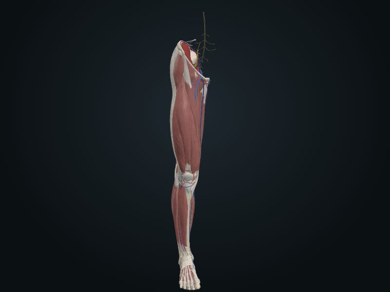
Study entry
Description
A downward extension of the medial side of the distal end; the lateral surface is articular.
Relations
It forms the medial wall of the ankle mortise and articulates with the talus; attachment of ligaments.
Palpation
Under the skin; blunt tip, about 1.25 cm above the tip of the lateral malleolus.
Source: cap. 7, «Huesos del miembro inferior: tibia» y «Anatomía de superficie»
Clinical correlation
Saphenous cutdown at the ankle and injury of the saphenous nerve
Even when it cannot be seen (infant, obese person, shock with collapsed veins), the great saphenous vein can always be found with a skin incision in front of the medial malleolus. This venous cutdown allows a cannula to be placed to give blood, volume expanders, electrolytes or drugs for a long time. The saphenous nerve accompanies the vein at this point; if it is cut or caught in the suture, the patient feels pain or numbness on the medial border of the foot.
Posterior tibial pulse and claudication
The pulse of the posterior tibial artery lies between the posterior surface of the medial malleolus and the medial border of the calcaneal tendon. Because the artery passes under the flexor retinaculum, ask the patient to invert the foot and relax the retinaculum; otherwise the pulse may seem absent. Compare both sides at the same time. It is essential in peripheral arterial occlusive disease: it is missing in 15% of normal young people, but its absence after 60 is a sign of the disease. An example is intermittent claudication, pain and cramp in the leg that appear on walking and pass with rest, from ischemia of the muscles.
Related clinical cases
- Saphenous cutdown at the ankle and injury of the saphenous nerve
- Posterior tibial pulse and claudication