 Espiral Atlas
Neck of the femur
collum femoris
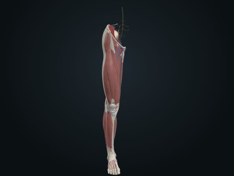
Study entry
Description
Trapezoid: the narrow end supports the head and the wide base continues with the shaft; with the shaft it forms the angle of inclination.
Source: cap. 7, «Huesos del miembro inferior: fémur»
Clinical correlation
Coxa vara and coxa valga
The angle of inclination between the neck and the shaft of the femur changes with age, sex and the development of the bone, and also with diseases that weaken the neck (rickets, for example). A smaller angle is coxa vara; a larger angle is coxa valga. «Varus» means that the distal part deviates toward the midline; «valgus», that it moves away from it. Coxa vara shortens the limb a little and limits passive abduction of the hip.
Slipped capital femoral epiphysis
Between 10 and 17 years of age, the epiphysis of the femoral head can slip on the neck because the epiphysial plate is the weak point. The cause is acute trauma or repeated microtrauma that shears the epiphysis, especially in abduction and lateral rotation of the thigh. The slip is usually slow and produces a progressive coxa vara. The first symptom is often discomfort in the hip that may be felt in the knee. A radiograph of the upper end of the femur confirms it.
Fractures of the femur
Although large and strong, the femur breaks often, and the type of fracture follows age and sex. That of the neck is the most common: it is the narrowest and weakest part and forms an angle with the line of load; it becomes more fragile with age, especially in women with osteoporosis. Proximal fractures (transcervical, intertrochanteric) come from indirect trauma, such as tripping or stepping down hard from a step. They are unstable: the fragments overlap and the limb shortens, helped by muscle spasm. Intracapsular ones also put the femoral head at risk, since it loses its blood supply.
Fracture of the neck of the femur and necrosis of the head
Calling it a «hip fracture» is improper, because it suggests the hip bone. It is rare in sport: before age 40 the neck is strong and only breaks with a lot of energy, with the limb extended (foot firm on the car floor, knee hitting the dashboard), and the force rises to the hip. After 60 it is very common, especially in women, because osteoporosis leaves the neck weak. It is usually intracapsular and needs internal fixation. The limb is rotated outward. Most of the blood of the head and neck comes from the medial circumflex femoral, through the retinacular arteries, which tear in the fracture of the neck and in dislocation of the hip.
Related clinical cases
- Coxa vara and coxa valga
- Slipped capital femoral epiphysis
- Fractures of the femur
- Fracture of the neck of the femur and necrosis of the head