 Espiral Atlas
Temporomandibular joint (TMJ)
articulatio temporomandibularis
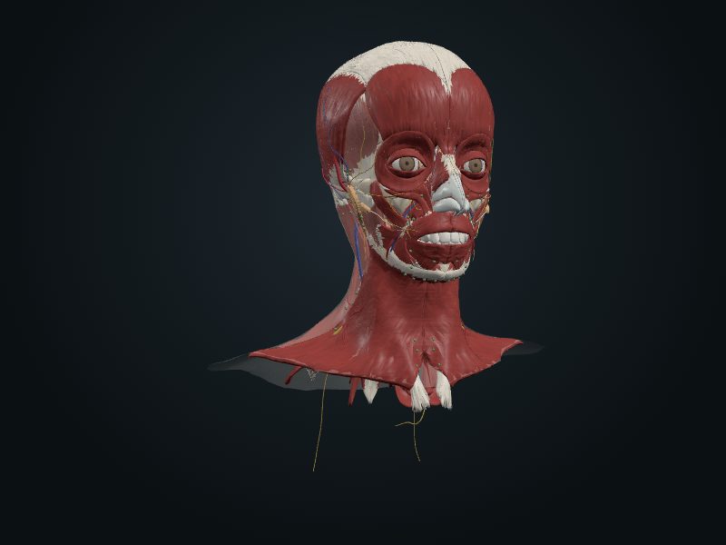
Study entry
Classification
Modified synovial hinge joint: besides the flexion (elevation) and extension (depression) typical of a hinge, it allows gliding (translation) and a small degree of rotation (spin).
Surfaces
The mandibular fossa and articular tubercle of the temporal bone, above, and the head of the mandible, below. An articular disc of fibrocartilage completely separates the two bony surfaces.
Capsule
A loose fibrous membrane, attached to the borders of the articular cartilage of the temporal bone and around the neck of the mandible. The disc, attached at its periphery to the inner surface of the capsule, creates two cavities, upper and lower, lined by upper and lower synovial membranes. A thicker part of the capsule forms the lateral ligament, which reinforces the joint laterally and, with the postglenoid tubercle, prevents posterior dislocation. Two extrinsic ligaments connect the mandible to the skull: the stylomandibular (thickening of the fibrous capsule of the parotid, from the styloid process to the angle of the mandible, not contributing to firmness) and the sphenomandibular (from the spine of the sphenoid to the lingula of the mandible), the main passive support of the mandible and a «swinging hinge» that serves as a fulcrum in the movements.
Movements
Protrusion and retrusion (translation) occur between the temporal bone and the disc (upper cavity); depression and elevation (hinge) and rotation, in the lower cavity. With the mouth closed, the heads of the mandible are retracted in the mandibular fossae. To open the mouth beyond what is needed to separate the dental arches, the head and disc move forward until they lie below the articular tubercle; only then is depression complete. If the advance is unilateral, the contralateral head rotates under the disc, which allows the simple grinding movements from side to side. The lateral pterygoid is the main mover of protrusion and depression; the temporalis (posterior fibers) and the masseter, of elevation and retrusion. Depression is usually done by gravity, and the suprahyoid and infrahyoid muscles act against resistance.
Blood supply
[CHECK: Moore does not describe it in the passages consulted (look in another source).]
Innervation
The auriculotemporal nerve supplies the articular branches.
Source: cap. 8, «Articulación temporomandibular» e tabla 8-10
Clinical correlation
Dislocation of the temporomandibular joint
On yawning or opening the mouth too wide for a big bite, excessive contraction of the lateral pterygoid muscles can dislocate the heads of the mandible forward, in front of the articular tubercles. The mandible is lowered and the mouth does not close. More commonly, however, a side punch to the chin, with the mouth open, dislocates the joint on the side of the blow. The dislocation can come with a fracture of the mandible. Posterior dislocation is rare, because of the postglenoid tubercle and the strong lateral ligament; in falls or direct blows to the chin, the neck of the mandible usually fractures first.
Related clinical cases
- Dislocation of the temporomandibular joint