 Espiral Atlas
Joints of the vertebral bodies (intervertebral discs)
symphyses intervertebrales
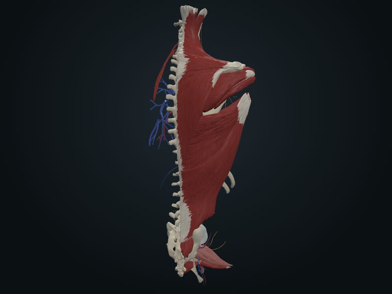
Study entry
Classification
Symphyses (secondary cartilaginous joints), designed to bear weight and give stability. The discs form the lower half of the anterior border of the intervertebral foramen.
Surfaces
Articular surfaces of the bodies of adjacent vertebrae, joined by discs. There is no disc between C1 and C2; the lowest functional disc is L5–S1.
Capsule
Each disc has an outer annulus fibrosus, made of concentric lamellae of fibrocartilage attached to the epiphysial rims of the bodies, with oblique fibers crossing in opposite directions (they allow limited rotation and give a strong union); the annulus is thinner behind. In the center lies the nucleus pulposus, gelatinous, about 88% water at birth, avascular (nourished by diffusion) and located between the center and the posterior part of the disc. The discs make up 20 to 25% of the height of the spine and are thicker in front in the cervical and lumbar regions, which creates the secondary curvatures.
Movements
They absorb shocks through their elastic deformability; the nucleus widens under compression and is the semi-liquid fulcrum in flexion, extension, lateral flexion and rotation. They account for most of the mobility of the spine.
Blood supply
[CHECK: Moore does not describe it in the passages consulted (look in another source).]
Innervation
Meningeal (recurrent) branches of the spinal nerves, distributed over the anterolateral surface of the vertebral bodies and the discs; they supply mainly the annuli fibrosi and the anterior longitudinal ligament.
Clinical
The posterior longitudinal ligament helps prevent or redirect posterior herniation of the nucleus pulposus.
Source: cap. 2, «Articulaciones de la columna vertebral»: articulaciones de los cuerpos vertebrales; «Vascularización de la columna vertebral» e «Nervios de la columna vertebral»