 Espiral Atlas
Knee joint
articulatio genus
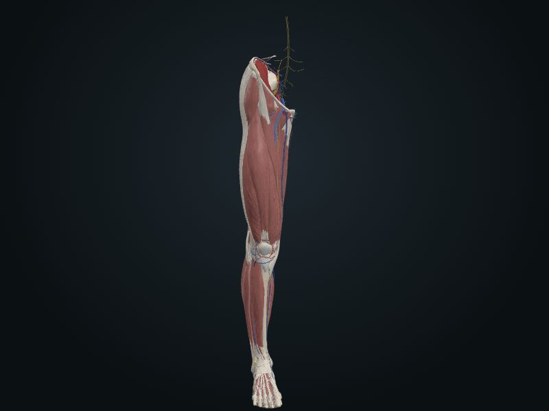
Study entry
Classification
The largest and most superficial in the body: largely a synovial hinge joint (flexion and extension with sliding, rolling and rotation). It consists of three joints: two femorotibial (lateral and medial) and one intermediate femoropatellar. The fibula does not take part.
Surfaces
Condyles of the femur and tibia (large, of complex and incongruent shapes, compared to two balls on a curved board), with the patellar surface of the femur and the patella. The medial meniscus (C-shaped, wider behind, attached to the tibial collateral ligament) and the lateral (almost circular, smaller and more mobile) deepen the tibial surface; the transverse ligament of the knee joins their anterior borders.
Capsule
Mechanically weak because of the incongruence; stability comes from the muscles (above all the quadriceps, especially the inferior fibers of the vastus medialis and lateralis) and from the ligaments. The capsule has an outer fibrous membrane, thin and incomplete, and an inner synovial one, which forms the suprapatellar bursa (5 cm above the patella), the alar folds and the infrapatellar fold. Five extracapsular ligaments: the patellar (the distal part of the quadriceps tendon, from the patella to the tibial tuberosity, with the medial and lateral patellar retinacula), the fibular collateral (extracapsular cord, from the lateral epicondyle to the head of the fibula; the popliteus passes under it), the tibial collateral (flat intrinsic band, from the medial epicondyle to the medial condyle of the tibia, fixed to the medial meniscus; it is injured more often), the oblique popliteal (expansion of the semimembranosus) and the arcuate popliteal. Intra-articular: the anterior (weaker) and posterior (stronger, from the back of the medial condyle) cruciate ligaments, which cross each other inside the capsule but outside the synovial cavity, and the menisci; the posterior meniscofemoral ligament connects the lateral meniscus to the posterior cruciate. The popliteus tendon passes through the capsule.
Movements
Flexion and extension (extension at 0°, flexion up to 120° with the hip extended and 140° with it flexed); rotation with the knee flexed (about 10° medial and up to 60° lateral at 90° of flexion). In full extension the knee «locks» by medial rotation of the femoral condyles; the popliteus unlocks it, rotating the femur laterally by 5°. The anterior cruciate prevents posterior displacement of the femur on the tibia and hyperextension; the posterior prevents anterior displacement of the femur and hyperflexion.
Blood supply
Articular arterial network with 10 vessels: genicular branches of the femoral and popliteal arteries and anterior and posterior recurrent branches of the anterior tibial recurrent and circumflex fibular arteries. The middle genicular artery of the popliteal pierces the capsule and supplies the cruciate ligaments, the synovium and the condyles.
Innervation
By Hilton's law, articular branches: anterior surface, femoral nerve (branches to the vasti); posterior, tibial nerve; lateral, common fibular nerve; also cutaneous branches of the obturator.
Clinical
The tibial collateral ligament and the medial meniscus tear frequently in contact sports.
Source: cap. 7, «Articulaciones del miembro inferior»: articulação da rodilla; tablas 7-16 e 7-17
Clinical correlation
Hip and knee arthroplasty
The hip is strong, but suffers severe trauma and wear. Osteoarthritis of the hip (pain, swelling, limitation, erosion of cartilage) is a common cause of disability. In arthroplasty, the head and neck of the femur are replaced by a metal prosthesis cemented into the femur, and a plastic cup is cemented into the hip bone in place of the acetabulum. In a very diseased knee (osteoarthritis, for example), total arthroplasty removes the defective parts and cements metal and plastic components onto the ends of the femur and tibia, imitating cartilage gliding on cartilage. It works well in people with a more sedentary life.
Genu varum and genu valgum; Q angle
The femur descends diagonally and the tibia is almost vertical, and the two form an angle at the knee. The Q angle measures it: a line from the anterior superior iliac spine to the center of the patella and another from the center of the patella to the tibial tuberosity. It is greater in women, with a wider pelvis. Normally the center of the knee lies under the head of the femur and the weight passes through the intercondylar region. In genu varum («bowlegs») the femur is too vertical, the Q angle decreases and the weight passes inside the knee: the medial surface suffers osteoarthritis and the fibular collateral ligament is overloaded.
Knee ligament injuries: the triad and drawer signs
The knee is injured often: it is low, mobile, bears weight and serves as the pivot between two long levers (thigh and leg), and its stability depends almost only on ligaments and muscles. In contact sport, the most common injury is the ligament sprain with the foot fixed on the ground. The tibial and fibular collaterals are taut with the knee extended.
Meniscal injuries
The meniscus that tears most is the medial; the lateral escapes because it is more mobile. Pain on rotating the tibia outward on the femur points to the lateral meniscus; on rotating inward, to the medial. Meniscal injuries usually come with rupture of the tibial collateral or the anterior cruciate. Tears at the periphery, which is well supplied with blood, can be repaired or heal on their own; those that do not heal are usually removed by arthroscopy.
Related clinical cases
- Hip and knee arthroplasty
- Genu varum and genu valgum; Q angle
- Knee ligament injuries: the triad and drawer signs
- Meniscal injuries
- Knee arthroscopy