 Espiral Atlas
Scapula
scapula
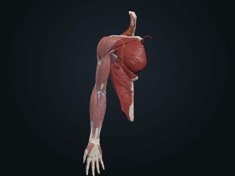
Study entry
Parts
Costal surface with the subscapular fossa; posterior surface divided by the spine into the supraspinous and infraspinous fossae; spine ending in the acromion; coracoid process; glenoid cavity with supraglenoid and infraglenoid tubercles; medial, lateral and superior borders (with the scapular notch); superior, inferior and lateral angles.
Joints
Glenohumeral (glenoid cavity with the head of the humerus) and acromioclavicular (acromion with the clavicle); the scapulothoracic is a gliding plane, not a true joint.
Palpation
Spine, acromion, inferior angle, medial border and tip of the coracoid process.
Clinical
Fractures are rare and high-energy. The scapular notch, closed by the superior transverse ligament, is where the suprascapular nerve may be compressed.
Source: cap. 3, «Huesos del miembro superior: escápula»
Clinical correlation
Fracture of the scapula
It requires strong trauma (being hit by a vehicle, for example) and usually comes with rib fractures. Because the scapula is covered with muscle on both sides, it almost always needs little treatment. The part that breaks most is the acromion, which lies under the skin.
Arterial anastomoses around the scapula
Around the scapula, the dorsal scapular, suprascapular and subscapular arteries (through the circumflex scapular branch) form networks on both surfaces of the bone. This collateral circulation saves the limb when the subclavian or an injured axillary must be ligated, or when the axillary narrows gradually from atherosclerosis. If the axillary is ligated between the 1st rib and the origin of the subscapular, blood flows backward in the subscapular and reaches the 3rd part of the axillary.
Related clinical cases
- Fracture of the scapula
- Arterial anastomoses around the scapula