 Espiral Atlas
Elbow joint
articulatio cubiti
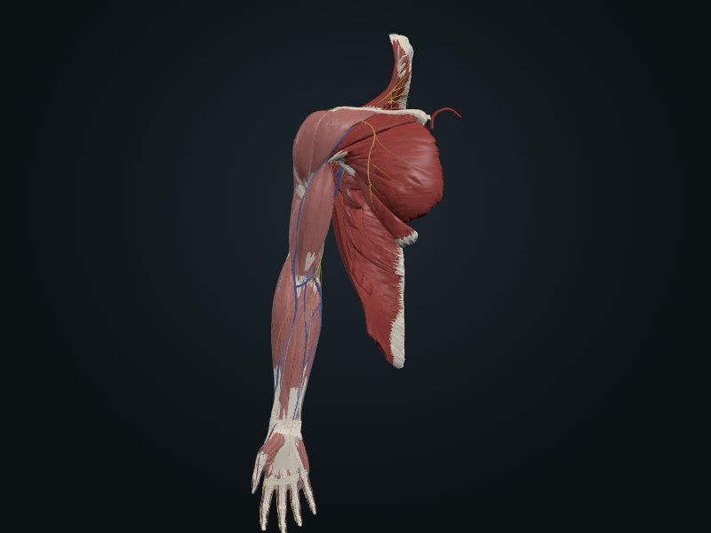
Study entry
Classification
Hinge synovial joint, 2–3 cm below the epicondyles.
Surfaces
Trochlea of the humerus with the trochlear notch of the ulna (humeroulnar) and capitulum of the humerus with the superior surface of the head of the radius (humeroradial); hyaline cartilage.
Capsule
Weak capsule in front and behind, attached to the humerus above the coronoid and olecranon fossae. Reinforced on the sides by the collateral ligaments: radial (fan-shaped, from the lateral epicondyle to the annular ligament) and ulnar (from the medial epicondyle to the coronoid process and the olecranon, with anterior, the strongest, posterior and oblique bands).
Movements
Flexion and extension. In extension, the axis of the ulna makes about 170° with that of the humerus (carrying angle), more acute in women. Main flexors: brachialis and biceps; main extensor: triceps.
Blood supply
Elbow arterial network (arterial anastomoses around the joint).
Innervation
Musculocutaneous, radial and ulnar nerves.
Clinical
Olecranon bursae: the subcutaneous one becomes inflamed from friction or a fall (bursitis). In children, forced abduction can tear off the medial epicondyle and injure the ulnar nerve. Rupture of the ulnar collateral ligament in pitchers is reconstructed with a tendon graft (Tommy John surgery).
Source: cap. 3, «Articulaciones del miembro superior: articulación del codo» y cuadro clínico
Clinical correlation
Avulsion of the medial epicondyle
In a child, a fall that forces abduction of the extended elbow makes the ulnar collateral ligament tear the medial epicondyle off downward. This is possible because the epiphysis of the medial epicondyle may only fuse to the humerus by 20 (on radiograph, generally at 14 in girls and 16 in boys). A common complication: stretching of the ulnar nerve, which passes behind the epicondyle.
Reconstruction of the ulnar collateral ligament ("Tommy John")
Ruptures and sprains of the ulnar collateral ligament are increasingly common in throwers (baseball, rugby, javelin, water polo). The reconstruction uses a long tendon from the person themself, from the forearm of the other side or from the leg (palmaris longus or plantaris): 10 to 15 cm of tendon pass through holes in the medial epicondyle of the humerus and in the side of the coronoid process of the ulna.
Dislocation of the elbow
Posterior dislocation happens when the child falls on the hands with the elbows flexed; it also comes from hyperextension or from a blow that pushes the ulna backward. With the radius and ulna behind, the end of the humerus ruptures the weak anterior part of the capsule. The ulnar collateral ligament usually tears, and there may be a fracture of the head of the radius, the coronoid process or the olecranon. If the ulnar nerve is injured, the little finger becomes numb and flexion and adduction of the wrist weaken.
Related clinical cases
- Avulsion of the medial epicondyle
- Reconstruction of the ulnar collateral ligament ("Tommy John")
- Dislocation of the elbow