 Espiral Atlas
Radiocarpal joint
articulatio radiocarpalis
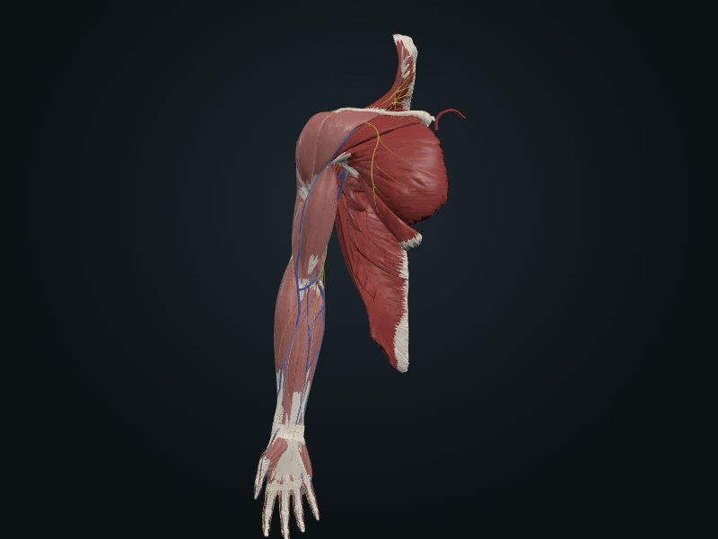
Study entry
Classification
Ellipsoid (condyloid) synovial joint. Its position is given by the line between the styloid processes of the radius and the ulna.
Surfaces
Distal end of the radius and articular disc of the distal radioulnar joint with the proximal row of the carpus (scaphoid, lunate and triquetrum); the ulna does not take part and the pisiform lies in front.
Capsule
Capsule attached to the radius, the ulna and the proximal row. Reinforcements: palmar and dorsal radiocarpal ligaments (oriented so that the hand follows the radius in supination) and the ulnar (styloid of the ulna–triquetrum) and radial (styloid of the radius–scaphoid) collaterals.
Movements
Flexion and extension (more flexion than extension), abduction and adduction (radial and ulnar deviation; more adduction) and circumduction, added to small glides in the intercarpal joints. Abduction is limited to about 15° by the styloid process of the radius.
Blood supply
Branches of the dorsal and palmar carpal arches.
Innervation
Anterior interosseous branch of the median, posterior interosseous branch of the radial, and dorsal and deep branches of the ulnar.
Clinical
The slightly extended position is the most stable and the functional resting position.
Source: cap. 3, «Articulaciones del miembro superior: articulación radiocarpiana»
Clinical correlation
Fractures of the radius and ulna; Colles fracture
In children they are usually greenstick fractures. A direct blow breaks both bones at the same level, generally in the middle third. Because the interosseous membrane joins the two, a fracture of one bone tends to come with dislocation of the nearest joint. The Colles fracture is the most common of the forearm: a complete transverse fracture of the distal 2 cm of the radius, frequent after 50 and in women with osteoporosis, from a fall on the outstretched, pronated hand. The distal fragment (often comminuted) goes backward, the styloid process of the ulna may be torn off and, with the radius shortened, the styloid of the radius is no longer more distal than that of the ulna.
Fracture of the scaphoid
It is the carpal bone that breaks most often, in a fall on the palm with the hand abducted; the line passes through the narrow part (waist). It hurts on the lateral side of the wrist, above all on extending and abducting the hand. The first radiograph may be normal; 10 to 14 days later, bone resorption shows the line. The proximal part is poorly supplied with blood: union may take 3 months or more, and the proximal fragment may become necrotic (avascular necrosis), leading to osteoarthritis of the carpus and, sometimes, arthrodesis.
Synovial cyst of the wrist (ganglion)
Sometimes a painless cystic lump appears, almost always on the dorsum of the wrist, the size of a small grape (it may reach that of a plum), with a thin wall and clear, viscous fluid. The cause is not known (perhaps mucoid degeneration). It grows and may hurt on flexing the wrist. It lies next to the synovial sheaths of the dorsum of the wrist and often communicates with them; another common site is the attachment of the extensor carpi radialis brevis at the base of the 3rd metacarpal. On the front of the wrist, a cyst of the common flexor sheath may narrow the carpal tunnel and compress the median nerve (carpal tunnel syndrome).
Related clinical cases
- Fractures of the radius and ulna; Colles fracture
- Fracture of the scaphoid
- Synovial cyst of the wrist (ganglion)