 Espiral Atlas
Femur
femur
Study entry
Parts
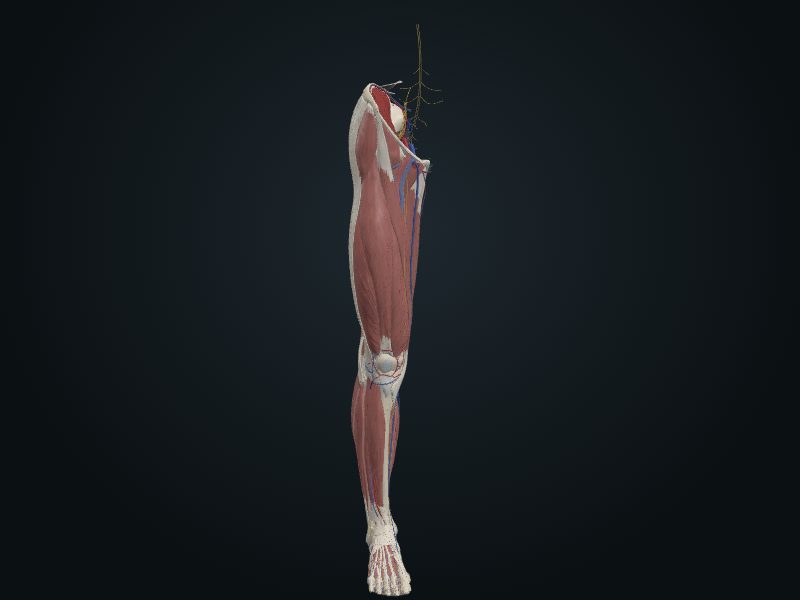
The longest and heaviest bone in the body (about a quarter of the height). Proximal end: head, neck and greater and lesser trochanters; the neck forms with the shaft the angle of inclination (115°–140° in the adult, average 126°, smaller in women) and with the axis of the condyles the angle of torsion (about 7° in men and 12° in women). Shaft: slightly convex forward, with the linea aspera behind. Distal end: medial and lateral condyles, intercondylar fossa and patellar surface.
Joints
Hip (head with the acetabulum); knee (condyles with the menisci and the condyles of the tibia; patellar surface with the patella).
Palpation
Only the ends: the greater trochanter, on the lateral surface of the thigh, about 10 cm below the iliac crest; the condyles and epicondyles, under the skin, at the knee; the adductor tubercle, at the top of the medial condyle. The head lies deep, a finger's breadth below the middle of the inguinal ligament.
Clinical
The angle of inclination puts a lot of load on the neck: in an older person with osteoporosis, a slight stumble can fracture the neck of the femur.
Source: cap. 7, «Huesos del miembro inferior: fémur» y «Anatomía de superficie de la cintura pélvica y el fémur»
Clinical correlation
Lower limb injuries in adolescents
Most lower limb injuries occur at the knee, leg and foot; the hip accounts for less than 3%. They come from acute trauma in contact sports or from overuse in endurance sports, such as the marathon. Adolescents are the most exposed: endochondral ossification only ends in early adulthood, and the epiphysial plates (discs of cartilage between metaphysis and epiphysis, which let the bone grow in length) are still open just when they train the most. During the growth spurt, bone grows faster than the muscles attached to it. The sum of effort and growth irritates and injures the plate and the developing bone (osteochondrosis).
Coxa vara and coxa valga
The angle of inclination between the neck and the shaft of the femur changes with age, sex and the development of the bone, and also with diseases that weaken the neck (rickets, for example). A smaller angle is coxa vara; a larger angle is coxa valga. «Varus» means that the distal part deviates toward the midline; «valgus», that it moves away from it. Coxa vara shortens the limb a little and limits passive abduction of the hip.
Fractures of the femur
Although large and strong, the femur breaks often, and the type of fracture follows age and sex. That of the neck is the most common: it is the narrowest and weakest part and forms an angle with the line of load; it becomes more fragile with age, especially in women with osteoporosis. Proximal fractures (transcervical, intertrochanteric) come from indirect trauma, such as tripping or stepping down hard from a step. They are unstable: the fragments overlap and the limb shortens, helped by muscle spasm. Intracapsular ones also put the femoral head at risk, since it loses its blood supply.
Genu varum and genu valgum; Q angle
The femur descends diagonally and the tibia is almost vertical, and the two form an angle at the knee. The Q angle measures it: a line from the anterior superior iliac spine to the center of the patella and another from the center of the patella to the tibial tuberosity. It is greater in women, with a wider pelvis. Normally the center of the knee lies under the head of the femur and the weight passes through the intercondylar region. In genu varum («bowlegs») the femur is too vertical, the Q angle decreases and the weight passes inside the knee: the medial surface suffers osteoarthritis and the fibular collateral ligament is overloaded.
Related clinical cases
- Lower limb injuries in adolescents
- Coxa vara and coxa valga
- Fractures of the femur
- Genu varum and genu valgum; Q angle