 Espiral Atlas
Tibia
tibia
Study entry
Parts
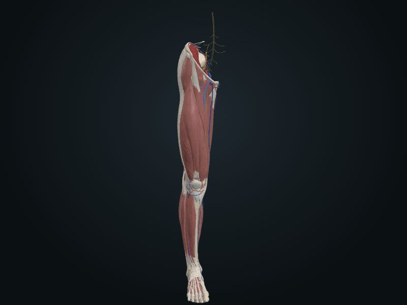
The second largest bone in the body, on the anteromedial side of the leg; it transmits the weight from the femur to the talus. Wide proximal end, with the medial and lateral condyles and the superior articular surface (the plateau), divided by the intercondylar eminence. Vertical shaft with a triangular cross-section, with three surfaces and three borders; thinnest at the junction of the middle and distal thirds. Smaller distal end, which extends downward on the medial side as the medial malleolus.
Joints
Knee (condyles with the condyles of the femur and the menisci); proximal tibiofibular (fibular articular facet with the head of the fibula); tibiofibular syndesmosis (fibular notch and interosseous membrane); talocrural (inferior surface and medial malleolus with the talus).
Palpation
Tibial tuberosity, about 5 cm below the apex of the patella; the medial surface and the anterior border (the shin), under the skin along their whole length; the condyles, on the sides of the patellar ligament; the medial malleolus, at the ankle.
Clinical
The anterior border and the medial surface lie just under the skin: the periosteum and skin of the shin are easily bruised.
Source: cap. 7, «Huesos del miembro inferior: tibia» y «Anatomía de superficie de la tibia y la fíbula»
Clinical correlation
Lower limb injuries in adolescents
Most lower limb injuries occur at the knee, leg and foot; the hip accounts for less than 3%. They come from acute trauma in contact sports or from overuse in endurance sports, such as the marathon. Adolescents are the most exposed: endochondral ossification only ends in early adulthood, and the epiphysial plates (discs of cartilage between metaphysis and epiphysis, which let the bone grow in length) are still open just when they train the most. During the growth spurt, bone grows faster than the muscles attached to it. The sum of effort and growth irritates and injures the plate and the developing bone (osteochondrosis).
Fractures of the tibia
The shaft of the tibia is narrowest at the junction of the middle and lower thirds: this is where it breaks most, and it is also the worst-supplied part. Because the anterior surface lies just under the skin, the tibia is the most common site of open fracture, for example when a car bumper strikes the leg. If the fracture passes through the nutrient canal and injures the nutrient artery, the fragments tend not to heal. Long untrained walks cause a stress fracture, transverse, in the lower third. Twisting with the foot fixed also breaks the tibia; in skiing, the twist gives a diagonal fracture at the middle-lower junction, often with the fibula, and the limb shortens.
Epiphysial plates of the tibia and Osgood-Schlatter disease
The center of ossification of the upper end of the tibia appears soon after birth and fuses with the shaft in adolescence, usually between 16 and 18 years. In a child, a fracture that crosses the epiphysial plate is more serious because it can interfere with growth. The tibial tuberosity usually grows downward from the upper center at about 10 years, but sometimes has a center of its own, near 12. If the plate is torn at the tuberosity, it becomes inflamed and hurts chronically and recurrently in adolescence: this is Osgood-Schlatter disease, typical of the young athlete.
Intraosseous infusion in the tibia
Intraosseous infusion delivers fluids, blood and drugs straight to the medullary cavity when a peripheral vein cannot be obtained, especially in traumatic shock and in a child in circulatory collapse. The most used site is the proximal tibia: the skin is thin and there are landmarks that help place the needle in the marrow without reaching the growth plate. The needle enters the flat area of the bone, about 2 cm below and slightly medial to the tibial tuberosity. Other sites: distal femur, distal tibia or fibula, proximal humerus and manubrium. Because of the risk of osteomyelitis, it is replaced by venous access within 24 hours.
Related clinical cases
- Lower limb injuries in adolescents
- Fractures of the tibia
- Epiphysial plates of the tibia and Osgood-Schlatter disease
- Intraosseous infusion in the tibia
- Tibial stress syndrome (shin splints)