 Espiral Atlas
Patella
patella
Study entry
Parts
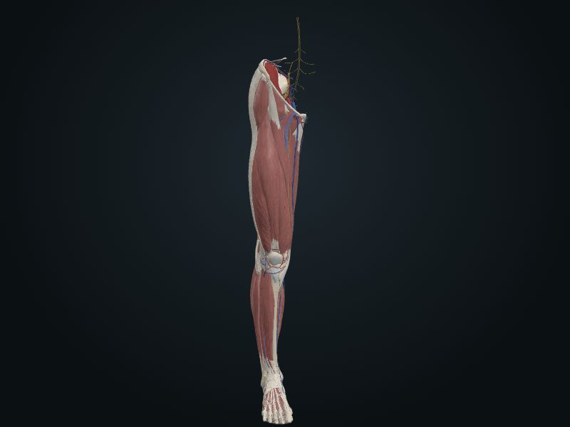
The largest sesamoid bone in the body, triangular, formed after birth within the tendon of the quadriceps femoris. Convex, subcutaneous anterior surface; thick base (upper margin); medial and lateral margins that descend to the apex; articular posterior surface covered by thick cartilage and divided by a vertical ridge into a medial part, narrower, and a lateral part, wider.
Joints
Femoropatellar, part of the knee (posterior surface with the patellar surface of the femur).
Palpation
The whole anterior surface, under the skin; it glides over the patellar surface of the femur when the knee flexes and extends.
Source: cap. 7, «Huesos del miembro inferior: patela» y «Anatomía de superficie de la cintura pélvica y el fémur»
Clinical correlation
Chondromalacia patellae and patellofemoral syndrome
Chondromalacia patellae is softening of the cartilage of the patella («runner's knee»), common in marathon runners and in running sports such as basketball. Pain around or behind the patella comes from imbalance of the quadriceps; it can also appear after a blow to the patella or extreme flexion of the knee (squatting to lift weight). In patellofemoral syndrome, the patella glides abnormally on the patellar surface of the femur and the repeated microtrauma hurts; direct contusion or osteoarthritis of this compartment also causes it.
Fracture of the patella
A direct blow can break the patella into two or more fragments. A transverse fracture comes from a blow to the knee or a sudden contraction of the quadriceps, as when slipping and trying not to fall backward. The upper fragment rises, pulled by the quadriceps tendon; the lower one stays attached to the patellar ligament.
Bipartite patella
At birth, the patella is cartilage. It ossifies between 3 and 6 years, often from more than one center. If the centers do not fuse, a bipartite or tripartite patella remains, which on a radiograph or CT can look like a fracture. It is almost always bilateral: examine both knees; if it is on both sides, it is probably an ossification variant.
Patellar reflex
With the patient seated and the legs hanging, a firm tap of the hammer on the patellar ligament extends the leg; a hand on the quadriceps feels the contraction. It is a deep (myotatic) reflex that tests the femoral nerve and segments L2–L4. The tap stretches the muscle spindles of the quadriceps; the impulse rises through the femoral nerve to L2–L4 and returns through the motor fibers of the same nerve, contracting the muscle. A diminished or absent reflex indicates injury at any point of the supply of the quadriceps, as in a peripheral neuropathy.
Related clinical cases
- Chondromalacia patellae and patellofemoral syndrome
- Fracture of the patella
- Bipartite patella
- Patellar reflex
- Dislocation of the patella